Archive for the ‘Exercise’ Category

Scientists have shown that when either lean or obese individuals exercise after eating a high fat meal, their fats are broken down and oxidized in skeletal muscle, making them healthier. These results show for the first time how a high fat diet and exercise stimulate the breakdown of fats and may help design ways to reduce excessive fat in the body.

Fat is broken down inside fat cells to generate energy by a process called lipolysis. The resulting fatty acids are released into the bloodstream and carried to tissues that require energy. In obese individuals, too much fat accumulates, compromising lipolysis, but the details of how this happens are not well understood. Also, obese individuals can show altered responsiveness to the stress hormones epinephrine and norepinephrine in their subcutaneous fat.

Max Lafontan and colleagues investigated how fat is broken down in both lean and obese subjects who exercised after either fasting or eating a high-fat diet. They noticed that after eating a high-fat diet, fats were broken down in both lean and obese individuals. Under fasting conditions, the breakdown of fats was more pronounced in the lean subjects, but the high fat meal enhanced lipolysis in the obese subjects.

The scientists also studied the effects of long-chain fatty acids (LCFAs) – which are found in high fat diet – on cultured fat cells. They noticed that LCFAs increase lipolysis when it is induced by epinephrine, one of the hormones known to stimulate lipolysis.

By showing for the first time how a high fat diet and LCFAs affect hormone-induced lipolysis in fat cells, this study paves the way for further research on the role of various fatty acids on the metabolism of muscle and blood vessel cells, the researchers conclude.

———————————–
Article adapted by MD Only from original press release.
———————————–

Article: “Acute exposure to long-chain fatty acids impairs alpha2-adrenergic receptor-mediated antilipolysis in human adipose tissue,” by Jan Polak, Cedric Moro, David Bessiere, Jindra Hejnova, Marie A. Marques, Magda Bajzova, Max Lafontan, Francois Crampes, Michel Berlan, and Vladimir Stich

MEDIA CONTACT: Max Lafontan, Institut National de la Sante et de la Recherche Medicale (French National Institute of Health and Medical Research), Toulouse, France; e-mail: Max.Lafontan@toulouse.inserm.fr

Research from Duke University Medical Center shows that even a modest amount of brisk walking weekly is enough to trim waistlines and cut the risk of metabolic syndrome (MetS), an increasingly frequent condition linked to obesity and a sedentary lifestyle.

It’s estimated that about a quarter of all U.S. adults have MetS, a cluster of risk factors associated with greater likelihood of developing heart disease, diabetes and stroke: large waist circumference, high blood pressure, high levels of triglycerides, low amounts of HDL, or “good” cholesterol, and high blood sugar. To be diagnosed with MetS, patients must have at least three of these five risk factors, and according to many studies, a growing number of people do.

But Johanna Johnson, a clinical researcher at Duke Medical Center and the lead author of a new study examining the impact of exercise on MetS, said a person can lower risk of MetS by walking just 30 minutes a day, six days per week. “That’s about 11 miles per week. And our study shows that you’ll benefit even if you don’t make any dietary changes.”

“The results of our study underscore what we have known for a long time,” said Duke cardiologist William Kraus. “Some exercise is better than none; more exercise is generally better than less, and no exercise can be disastrous.”

The study appears in the December 15 issue of the American Journal of Cardiology.

The results come from a multi-year, federally funded study called STRRIDE (Studies of a Targeted Risk Reduction Intervention through Defined Exercise) that examined the effects of varying amounts and intensity of exercise on 171 middle-aged, overweight men and women.

Before exercising regularly, 41 percent of the participants met the criteria for MetS. At the end of the 8-month exercise program, only 27 percent did.

“That’s a significant decline in prevalence,” said Johnson. “It’s also encouraging news for sedentary, middle-aged adults who want to improve their health. It means they don’t have to go out running four or five days a week; they can get significant health benefits by simply walking around the neighborhood after dinner every night.”

Still, some exercise regimens were better than others. Those who exercised the least, walking about 11 miles per week, gained significant benefit, while those who exercised the most, jogging about 17 miles per week, gained slightly more benefit in terms of lowered MetS scores.

One group puzzled the researchers, however. Those who did a short period of very vigorous exercise didn’t improve their MetS scores as much as those who performed less intense exercise a longer period.

Kraus said there may be more value in doing moderate intensity exercise every day rather than more intense activity just a few days a week.

In all three of the study’s exercise groups, waistlines got smaller over the 8-month period. In general, men who exercised saw greater improvement in their MetS risk factors than women. But Johnson points out that at baseline, the men generally had worse scores than women, “so they had more room to improve,” she said.

Over the course of the STRRIDE study, the inactive control group – those who didn’t change their diet or activity level at all – gained an average of about one pound and a half-inch around the waist. “That may not sound like much, but that’s just six months,” Kraus said. “Over a decade, that’s an additional 20 pounds and 10 inches at the beltline.”

—————————-
Article adapted by MD Only Weblog from original press release.
—————————-

Contact: Michelle Gailiun
Duke University Medical Center

The study was funded by the National Institutes of Health.

Colleagues at Duke who contributed to the study include Cris Slentz, Gregory Samsa, Lori Bateman and Brian Duscha. Collaborating authors from East Carolina University include Joseph Houmard, Jennifer McCartney and Charles Tanner.

Diabetes is the leading cause of blindness among people aged 20-74 in Minnesota. Each year, according to the Minnesota Department of Health (MDH), between 400 and 700 Minnesotans go blind due to diabetes complications. Even when blood sugar is controlled, 60% of those having diabetes for more than 15 years will experience some form of diabetic eye disease in their lifetime.November is American Diabetes Month. This is a good time to schedule your annual dilated eye exam, urges the Minnesota Optometric Association (MOA), because type 2 diabetes can be caught early with this exam, and treated. MDH statistics show that of the 276,000 Minnesotans who have diabetes, 96,000 are unaware that they have it.

Currently, only eight in 10 Minnesotans with diabetes have a dilated eye exam every two years. “Most people do not realize that diabetics are twice as likely to suffer from cataracts, diabetic retinopathy and glaucoma. People may have these diseases before they are aware of it,” said Dr. Dirk Colby, president of the MOA and a doctor of optometry with West Metro Ophthalmology, Golden Valley and Plymouth, Minnesota. “It is critical for diabetic patients to come in every year,” said Dr. Colby. “Waiting two years or even worse, not coming in at all, can have a catastrophic effect.”

Diabetes is taking its toll on Americans and Minnesotans with eye diseases and type 2 diabetes is increasing rapidly due to the increase in obesity and decrease in exercise. These two risk factors are the leading causes of type 2 diabetes.

Diabetic retinopathy, the most common diabetic eye disease, is the leading cause of blindness in Minnesota, and overall in the United States. Diabetic retinopathy is caused by changes in the blood vessels of the retina. In some people with diabetic retinopathy, retinal blood vessels may swell and leak fluid; in others, abnormal new blood vessels grow on the surface of the retina. These changes may result in vision loss or blindness.

Cataracts occur when the normally clear lens in the eye becomes cloudy, impairing vision. Clouded vision may make it more difficult to read, drive, or see clearly. Glaucoma, an increase in fluid pressure inside the eye, leads to optic nerve damage and vision loss.

To help prevent diabetic eye diseases, the American Diabetes Association recommends people suffering from diabetes should:

  • Keep your blood sugar under good control. People who keep their blood sugar levels closer to normal are less likely to have retinopathy.
  • Bring high blood pressure down. High blood pressure can make eye problems worse.
  • Get a dilated eye exam by a family eye doctor every year. Your optometrist will use drops to enlarge your pupils to look inside your eyes.

The Minnesota Optometric Association (MOA) recommends you see your family eye doctor right way if you have any of the following problems:

  • Your vision is blurry.
  • You see double, spots or floaters
  • One or both eyes hurt
  • You feel pressure in your eye
  • You can’t see things at the periphery as well as before
  • You have trouble reading

—————————-
Article adapted by MD Only Weblog from original press release.
—————————-

Contact:
Jim Meffort-Nelson
minnesota.aoa.org

Perhaps George Washington wouldn’t have chopped down his father’s cherry tree if he knew what chemists now know. They have identified a group of naturally occurring chemicals abundant in cherries that could help lower blood sugar levels in people with diabetes. In early laboratory studies using animal pancreatic cells, the chemicals, called anthocyanins, increased insulin production by 50 percent, according to a peer-reviewed study scheduled to appear in the Jan. 5 issue of the American Chemical Society’s Journal of Agricultural and Food Chemistry. ACS is the world’s largest scientific society.Anthocyanins are a class of plant pigments responsible for the color of many fruits, including cherries. They also are potent antioxidants, highly active chemicals that have been increasingly associated with a variety of health benefits, including protection against heart disease and cancer.

“It is possible that consumption of cherries and other fruits containing these compounds [anthocyanins] could have a significant impact on insulin levels in humans,” says study leader Muralee Nair, Ph.D., a natural products chemist at Michigan State University in East Lansing. “We’re excited with the laboratory results so far, but more studies are needed.” Michigan is the top cherry producing state in the nation.

Until human studies are done on cherry anthocyanins, those with diabetes should continue following their doctor’s treatment recommendations, including any medicine prescribed, and monitor their insulin carefully, the researcher says. The compounds show promise for both the prevention of type 2 (non-insulin-dependent) diabetes, the most common type, and for helping control glucose levels in those who already have diabetes, he adds.

While fresh cherries and fruits containing these anthocyanins are readily available, medicinal products may be the most efficient way to provide the beneficial compounds, according to Nair. It’s possible that anthocyanins eventually could be incorporated into new products, such as pills or specialty juices that people could take to help treat diabetes. Such disease-specific products may take several more years to develop, he notes.

Scientists in Nair’s laboratory have even developed a unique process, patented by the university, for removing sugar from fruit extracts that contain anthocyanins. This could lead to “sugar-free” medicinal products for people with diabetes.

The current study, partially funded by the U.S. Department of Agriculture, involved tart cherries (also known as sour cherries or pie cherries), a popular variety in the United States, and the Cornelian cherry, which is widely consumed in Europe. Nair and his associates, B. Jayaprakasam, Ph.D., L.K. Olson, Ph.D., and graduate student S. K. Vareed, tested several types of anthocyanins extracted from these cherries against mouse pancreatic-beta cells, which normally produce insulin, in the presence of high concentrations of glucose.

Insulin is the protein produced by the pancreas that helps regulate blood sugar (glucose) levels. Compared to cells that were not exposed to anthocyanins, exposed cells were associated with a 50 percent increase in insulin levels, the researchers say. The mechanism of action by which these anthocyanins boost insulin production is not known, Nair says.

Nair and his colleagues are currently feeding anthocyanins to a group of obese, diabetic mice to determine how the chemicals influence insulin levels in live subjects. Results of these tests are not yet available.

Although other fruits, including red grapes, strawberries and blueberries, also contain anthocyanins, cherries appear to be the most promising source of these compounds on the basis of serving size, according to the researcher. The compounds are found in both sweet and sour (tart) cherry varieties.

The potential benefits of cherries extend beyond diabetes. Previous studies by the researcher found that certain anthocyanins isolated from cherries have anti-inflammatory properties and may be useful in fighting arthritis. Nair’s colleagues have found that cherries also may help fight colon cancer.

But people with diabetes are encouraged to use caution when it comes to consuming maraschino cherries, the bright red candied version that adorns ice cream and cocktails, Nair points out. Many of the beneficial cherry pigments that were present in the fresh fruit have been removed during processing, replaced with food coloring, and extra sugar has been added.

The American Chemical Society is a nonprofit organization, chartered by the U.S. Congress, with a multidisciplinary membership of more than 159,000 chemists and chemical engineers. It publishes numerous scientific journals and databases, convenes major research conferences and provides educational, science policy and career programs in chemistry. Its main offices are in Washington, D.C., and Columbus, Ohio.

—————————-
Article adapted by MD Only Weblog from original press release.
—————————-

Contact: Michael Bernstein
American Chemical Society

Research has shown that people who exercise do better on memory tests. Now a new Columbia University Medical Center study explains specifically what exercise does within the brain. Exercise, the researchers found, targets a region of the brain within the hippocampus, known as the dentate gyrus, which underlies normal age-related memory decline that begins around age 30 for most adults.

This finding is significant because it was accomplished via the first-ever observation of neurogenesis, the growth of neurons, within a living brain. Using an MRI imaging technique developed at Columbia, the researchers were able to identify neurogenesis within the dentate gyrus region following exercise. Previously, researchers were only able to prove neurogenesis upon postmortem exam in animal studies.

“No previous research has systematically examined the different regions of the hippocampus and identified which region is most affected by exercise,” said Scott A. Small, M.D., associate professor of neurology at Columbia University Medical Center and the study’s lead author. “I, like many physicians, already encourage my patients to get active and this adds yet another reason to the long list of reasons why exercise is good for overall health.”

Published in the March 12-16, 2007 early online edition of the Proceedings of the National Academy of Sciences, the finding builds upon previous research at Columbia that identified the role of the dentate gyrus region of the hippocampus in normal age-related memory decline. Additionally, Fred “Rusty” Gage, Ph.D. of the Salk Institute, a lead co-investigator on this study, had demonstrated in mice that the dentate gyrus is the one area of the brain where new neurons are generated, and that exercise improves this process. This is the first human study to emerge out of this observation.

“Our next step is to identify the exercise regimen that is most beneficial to improve cognition and reduce normal memory loss, so that physicians may be able to prescribe specific types of exercise to improve memory,” said Dr. Small, who is also a research scholar at the Columbia University Taub Institute for Research on Alzheimer’s Disease and the Aging Brain.

—————————-
Article adapted by MD Only Weblog from original press release.
—————————-  

Contact: Elizabeth Streich, Columbia University Medical Center

Additional Columbia researchers who contributed to this study include: Ana C. Pereira, Rene Hen, Dan E. Huddleston, Adam M. Brickman, Alexander A. Sosunov, Guy M. McKhann, Truman R. Brown and Richard Sloan.

The Taub Institute for Research on Alzheimer’s Disease and the Aging Brain at Columbia University Medical Center is a multidisciplinary group that has forged links between researchers and clinicians to uncover the causes of Alzheimer’s, Parkinson’s and other age-related brain diseases and discover ways to prevent and cure these diseases. It has partnered with the Gertrude H. Sergievsky Center at Columbia University Medical Center which was established by an endowment in 1977 to focus on diseases of the nervous system. The Center integrates traditional epidemiology with genetic analysis and clinical investigation to explore all phases of diseases of the nervous system.

Columbia University Medical Center provides international leadership in basic, pre-clinical and clinical research, in medical and health sciences education, and in patient care. The medical center trains future leaders and includes the dedicated work of many physicians, scientists, nurses, dentists, and public health professionals at the College of Physicians & Surgeons, the College of Dental Medicine, the School of Nursing, the Mailman School of Public Health, the biomedical departments of the Graduate School of Arts and Sciences, and allied research centers and institutions.

According to a new study published in Journal of Thrombosis and Haemostasis, regular participation in sports reduces the risk of developing blood clots by 39 percent in women and 22 percent in men.

Researchers from Leiden University Medical Center in the Netherlands evaluated 7,860 people aged 18-70. Patients who had suffered their first blood clot in a leg vein or lung artery were compared with control subjects who had never experienced blood clots. 31 percent of the patients and 40 percent of the control group participated in sports on a regular basis.

Overall figures for both sexes showed that participating in sports at least once per week, regardless of the type of sport or its intensity, reduced the risk of developing a blood clot in a lung artery by 46 percent and a blood clot in a leg vein by 24 percent.

“Women were shown to be even more likely to reap the benefits of regular sporting activities than men,” says F.R. Rosendaal, co-author of the study. “When we excluded women who were pregnant or receiving oral contraceptive or hormone replacement therapy – all possible causes of blood clots – the risk for women was reduced by 55 percent.”

The authors note that, while strenuous activity is known to increase the risk of blood clot development in the elderly, regular exercise is also shown to greatly benefit the heart, and that the net effect of elderly sports participation may be positive.

The findings also show that people who did not participate in sports were more than four-times as likely to develop a blood clot if they were obese (with a body mass index of 30 or greater) than lean (with a body mass index of less than 25).

“When we looked at the results, we found that, overall, the mere fact that people took part in a sporting activity at least once a week was enough to lower their risk of blood clots,” say the authors.

—————————-
Article adapted by MD Only Weblog from original press release.
—————————-  

Contact: Sean Wagner
http://www.blackwellpublishing.com

_________________________________________________________________
This study is published in Journal of Thrombosis and Haemostasis.

To view the abstract for this article, please click here.

Journal of Thrombosis and Haemostasis publishes high quality, original research reports, state-of-the art reviews, invited commentaries and debates on timely topics, letters and announcements on thrombosis, bleeding disorders and vascular biology. It is produced monthly by Wiley-Blackwell on behalf of the International Society on Thrombosis and Haemostasis.

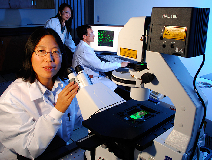
Liyun Wang, UD assistant professor of mechanical engineering, views osteocytes (bone cells) in her lab, as graduate students Wen Li and Xiaozhou Zhou examine the magnified images in the background. 

Click here for more information.


Ten million people in the United States are estimated to already have bone diseases, and almost 34 million more are estimated to have low bone mass, putting them at increased risk for osteoporosis, according to the National Osteoporosis Foundation.

Liyun Wang, assistant professor of mechanical engineering at the University of Delaware, knows the serious consequences of osteoporosis.

Two of Wang’s aunts have suffered from the insidious bone-thinning disease, and one aunt died within a year after falling and fracturing her hip.

Wang is now leading research that will shed light on how osteocytes–the cells encased inside your bones–sense external stimuli and communicate with cells on the surface, signaling them to either build more bone or remove existing bone.

The five-year, $1.6 million project, ranked in the top 5 percent of research proposals recommended for funding by the National Institutes of Health (NIH) this year, holds promise in unveiling the mysteries of bone and joint diseases afflicting people worldwide.

The results may not only help scientists home in on the cause of osteoporosis and arthritis, but also develop more effective drug therapies to treat the debilitating bone and joint diseases.

The project will involve an interdisciplinary team of investigators at UD, including Prof. Mary C. Farach-Carson and Associate Prof. Randall Duncan, who hold primary appointments in biological sciences with joint appointments in mechanical engineering, and John Novotny, assistant professor of mechanical engineering.

“Bone and joint disorders affect almost half of all people over 50 years old, at a cost of $250 billion annually in the United States,” Wang said. “A third of the people who suffer a fracture due to bone loss end up dying within a year.”

The embedded bone cells, or osteocytes, that Wang is studying, act like the bone’s “brain.”

“The osteocytes are very smart,” Wang says. “They can tell whether a person is using his or her bones or not. If the person is physically active, the osteocytes tell cells on the surface that it’s okay to put on more bone. Otherwise, they signal the surface cells to remove bone at a rate that can be as high as 3 percent bone mass per month, which is the case for patients confined to long-term bed rest and for astronauts,” she notes.

The osteocytes lie in tiny pits or holes, called lacunae, within the bone. These living cells have many long arms that connect them to surface bone cells and the bone’s vascular system. The narrow channels housing the osteocyte’s arms (canaliculi) and the lacunae form a network through which a mixture of water, nutrients and other bioactive molecules flows.

“Although it is hard as cement, bone is actually like a stiff sponge,” Wang says. “It’s porous and has water inside. When we have mechanical loading, when you run, for example, a part of the leg bone is compressed, and water is pushed through gaps, less than a micrometer in size, between the osteocytes and the bone cement that surrounds them.”

This powerful wave of fluid keeps the osteocytes happy and functioning well, Wang says, delivering nutrients to them from nearby blood vessels and quickly dispersing signaling molecules, such as calcium ions, from one cell to its neighbors.

Using a novel microscopic imaging method that Wang developed, which is based on fluorescence recovery after photobleaching (FRAP), the research team hopes to do what no one has done before: see inside living bone and determine how rapidly these signaling and nutrient molecules are transferred between the cells when a bone is at rest and when it is at work.

A high-powered laser-scanning microscope will be used to assess the movement of molecules in the tibia of an anesthetized mouse. A harmless green fluorescent dye, tagged to various-sized proteins, signaling molecules and cell nutrients, will be injected into the animal’s bloodstream.

The dyed molecules will be subjected to a flash of intense light, temporarily photobleaching them black, leaving a distinct black sector surrounded by green. Thus, if the molecules diffuse into one another’s territories, they can easily be tracked by color.

Using similar techniques, Wang is investigating the communication between bone and cartilage in the development and progression of osteoarthritis, one of five projects in an $11 million NIH grant led by Thomas Buchanan, professor and chairperson of the UD Department of Mechanical Engineering. The UD effort includes a unique mentoring program for women scientists.

Wang says she is grateful for the mentoring and support she has received from her colleagues at UD since she joined the faculty in 2005, as well as from her doctoral advisers at the City College of New York, including Profs. Susannah Fritton, Steve Cowin, and Sheldon Weinbaum; and Dr. Mitchell Schaffler, with whom she worked as a postdoctoral researcher at the Mt. Sinai School of Medicine.

Currently, Wang’s laboratory group includes Wen Li, a graduate student in biomechanics and movement science, Xiaozhou Zhou, a graduate student in mechanical engineering, and undergraduate students Ben Keller and Laura Schultz, who are both working on degrees in mechanical engineering, and Samantha Nigro, who is pursuing her degree in biological sciences. Research associate Jun Pan will join the group at the end of this year.

“My students have been excellent,” Wang says, smiling. “They are well-organized and eager to learn how to do research. They are very motivated, and that is important. We have exciting work ahead of us!”

—————————-
Article adapted by MD Only Weblog from original press release.
—————————-  

Contact: Tracey Bryant
tbryant@udel.edu
302-831-8185
University of Delaware

Wang is seeking two additional doctoral students and one postdoctoral researcher in the areas of biomechanics, orthopedic biology or mechanical engineering to join her research team. For more information, contact Wang at [lywang@udel.edu].

According to the National Osteoporosis Foundation, osteoporosis affects more than 2 million men in the United States and nearly 12 million more have osteopenia — clinically significant low bone density that is less severe than osteoporosis.

Now, a new study from the University of Missouri-Columbia has found that men engaging predominantly in low-impact forms of exercise have an increased incidence of osteopenia — a condition resulting in two times the risk of bone fracture.

Unfortunately, some individuals who believe they are doing everything right in terms of their health might be surprised and upset by our finding,” said Pamela Hinton, an associate professor of nutritional sciences in MU’s College of Human Environmental Sciences, who co-authored the study. “We believe, however, that these results will ultimately serve as education and motivation for these people.”

Hinton said the effects of osteopenia can be mitigated by integration of weight-bearing activities into the lifestyle of active individuals. Studies in pre- and post-menopausal women suggest that bone mineral density will increase 2 percent to 3 percent after six months of resistance training three times per week. Small changes in bone density translate into much larger changes in bone strength — a 1 percent increase in bone density reduces the risk of fracture by up to 5 percent.

“Regular, non-weight-bearing activities, such as swimming and cycling are effective measures for preventing the leading risk factors for death and disability in our society,” Hinton said. “But the results of this study suggest that regular weight-bearing activities, such as running, jogging, or rope jumping, are important for the maintenance of healthy bones.”

The researchers measured bone mineral density in 43 competitive male cyclists and runners ages 20 to 59. Findings of the study included:

* The cyclists had significantly lower bone mineral density of the whole body, especially of the lumbar spine, compared to runners.

* 63 percent of the cyclists had osteopenia of the spine or hip compared with 19 percent of the runners.

* Cyclists were seven-times more likely to have osteopenia of the spine than the runners. Background facts:

* The risk of fracture is increased approximately two-fold in osteopenic individuals and five-fold in people with osteopenia.

* Low bone density in males often remains undiagnosed and inadequately treated and, after suffering a fracture, men are less likely to receive follow-up care than women.

* Risk factors for osteoporosis in men are similar to those identified in women: family history, age, low body weight, smoking, excessive alcohol consumption, inadequate calcium or vitamin D intake, low reproductive hormone levels, physical inactivity, and disease or medication affecting bone metabolism.

—————————-
Article adapted by MD Only Weblog from original press release.
—————————-

The study, “Participation in road cycling versus running is associated with lower bone mineral density in men,” will be published in Metabolism, and is authored by MU researchers R.S. Rector, R. Rogers, M. Ruebel and P.S. Hinton, in the Department of Nutritional Sciences.

Source: Jennifer Faddis
University of Missouri-Columbia

Many young women are unaware of the dangers of osteoporosis, leaving them vulnerable to crippling bone disease.

Mark J. Kasper, Ed.D., FACSM was the lead researcher for a survey of 302 college females enrolled in an elective physical activity course. While almost all (97.7 percent) had heard of osteoporosis, only 52.9 percent were able to correctly identify eight or more out of 14 risk factors listed and only 30.8 percent named all three major risk factors: low calcium intake, a sedentary lifestyle, and infrequent menstruation. The National Osteoporosis Foundation defines osteoporosis as “a disease characterized by low bone mass and structural deterioration of bone tissue, leading to bone fragility and an increased susceptibility to fractures, especially of the hip, spine and wrist.”

Kasper pointed out that 39.7 percent of respondents believed they were unlikely to develop osteoporosis. “Despite a similar lifetime risk of developing osteoporosis, heart disease or breast cancer, there is a lack of knowledge and a high degree of complacency concerning osteoporosis in young women,” said Kasper. “This data mirrors our previous research suggesting that educational interventions targeting young women either have not been implemented or have been ineffective.”

The coeds in Kasper’s study that osteoporosis is a serious illness, but less serious than heart disease or breast cancer. Blacks were less likely than whites to identify risk factors and to believe that they would develop osteoporosis. Respondents ranged from 18 to 21 years of age.

Exercise Aids Bone HealthPhysical activity is the only known intervention that can potentially increase bone mass and strength in the early years of life and reduce the risk of falling in older populations, according to a Position Stand from the American College of Sports Medicine (ACSM). The official ACSM pronouncement, published in the November 2004 issue of Medicine & Science in Sports & Exercise®, encourages the adoption of specific exercise prescriptions designed for various ages to best capitalize on the chances to accrue and preserve bone throughout the various stages of life.

The paper cites worldwide predictions that the number of hip fractures will double by the year 2025, with a mortality rate of 15 to 20 percent within the first year following such a fracture for elderly individuals, as reasons to encourage a better understanding of the types, durations and frequencies of physical activities that help build and maintain bone. These include high-intensity, high-impact activities for children to build bone and moderate-to-high intensity weight-bearing activities for adults to maintain bone mass.

For adults, experts have established these recommendations to help preserve bone:

Mode – weight-bearing endurance activities such as tennis and jogging, activities that involve jumping, and resistance exercise (weight lifting)
Intensity – moderate to high
Frequency – weight-bearing activities 3 to 5 times per week, resistance exercise 2 to 3 times per week
Duration – 30 to 60 minutes a day combined

For children, especially in the years surrounding the onset of puberty, experts recommend the following activities to help build bone:

Mode – impact activities such as gymnastics and jumping activities combined with moderate resistance training
Intensity – high, but with appropriate weights for resistance training (no more than 60 percent of the maximum amount a person can lift one time)
Frequency – at least three days per week
Duration – 10 to 20 minutes with multiple sessions within the same day potentially being more effective

The Position Stand also explicitly mentions the importance of bone health for men, noting that the predicted increase in osteoporotic falls and fractures in men in the coming decades is even greater than in women. The authors also encourage weight-bearing physical activities for even the most frail and elderly, provided they can still perform them safely.

The conclusions outlined in this news release are those of the researchers only, and should not be construed as an official statement of the American College of Sports Medicine.

—————————-
Article adapted by MD Only Weblog from original press release.
—————————-

Contact: Communications and Public Information
American College of Sports Medicine

Some brains may be wired to encourage fidgeting and other restless behaviors that consume calories and help control weight, according to new research published by The American Physiological Society.

The study found that the brains of rats bred to be lean are more sensitive to a chemical produced in the brain, orexin A, which stimulates appetite and spontaneous physical activity such as fidgeting and other unconscious movements. Compared to rats bred to be obese, the lean rats had a far greater expression of orexin receptors in the hypothalamus.

“The greater expression of orexin receptors suggests the lean rats’ brains were more sensitive to the orexin the brain produces,” said Catherine M. Kotz, the study’s senior researcher. “The results point to a biological basis for being a couch potato.”

This line of research suggests that frequent minor unconscious movements such as fidgeting and other behaviors associated with restlessness burn calories and help control weight, Kotz said. Further, it suggests a strategy to reduce weight gain and could lead to the development of a drug to stimulate minor activity.

The study “Elevated hypothalamic orexin signaling, sensitivity to orexin A and spontaneous physical activity in obesity resistant rats,” appears in the online edition of the American Journal of Physiology-Regulatory, Integrative and Comparative Physiology published by The American Physiological Society. The study was done by Jennifer A. Teske and Allen S. Levine of the University of Minnesota and the Minnesota Obesity Center, St. Paul; Michael Kuskowski, VA Medical Center, Minneapolis; James A. Levine, Mayo Clinic, Rochester, Minnesota; and Catherine M. Kotz, the VA Medical Center, University of Minnesota, and the Minnesota Obesity Center.

Study looks at obese versus lean rats

“Many people focus on diet, but it may be more feasible for some people to stand or move more throughout the day” as a way to control their weight, Kotz said. Contrary to common belief, metabolism rates don’t vary greatly from person to person and weight gain usually results from eating too much, burning too few calories, or both, she said.

The researchers drew their conclusions after performing a series of experiments with obesity-prone and obesity-resistant rats. The obesity-prone strain was developed for obesity research by breeding obese rats with other obese rats. The obesity-resistant rats were developed by breeding lean rats with lean rats, Kotz noted. The study also employed a control group of normal laboratory rats.

Each rat consumed the same number of calories each day. The researchers took baseline measurements of each rat’s activity using sensors to measure even minor movements, such as grooming and standing.

They found that the lean group moved significantly more during this baseline period than the obese group, Kotz said. This was true even though the rats were young and both groups weighed the same — eliminating the obesity itself as the cause of the decreased movement. After the baseline data gathering, the researchers moved to the experimental part of the study.

Lean rats have elevated expression of orexin receptors

“We knew from previous studies that orexin stimulated physical activity, and so we wanted to find out whether it enhances activity more in lean rats than in obese rats, Kotz explained. The researchers injected orexin into the lateral hypothalamus area of the brains of both groups and found that the lean rats became even more active, while the obese rats didn’t respond much at all. “Not only do the lean rats have a higher base activity rate but they respond more to orexin,” she said.

Orexin must bind to receptors in the brain to produce increased activity, so the researchers reasoned that the lean rats must have more orexin receptors. When they did a blind analysis of the brains of obese and lean rats of various ages, they found that the lean rats had double the gene expression level of orexin receptors compared to the obese rats, Kotz explained.

The greater gene expression of orexin receptors does not conclusively prove that there are more orexin receptors, but it is highly suggestive of that finding. Kotz and her fellow researchers are now looking to see if the lean rats have a greater number of orexin receptors in their brains.

Activity level important to weight control

Because the rats in this study ate the same amount of food, the researchers concluded that the weight gain of the obese rats comes more from expending too few calories than from consuming too many. Other studies have shown that disabling the orexin system of lean rats causes them to eat less and move less, which leads them to become obese, Kotz said. When the orexin system is working optimally, the increase in eating which orexin causes is believed to be offset by increased physical activity, she said.

It would be impossible to do a similar study of the brain in humans. But one of the researchers, James Levine, found in a previous study with humans that lean individuals move about two hours per day more than obese individuals. What does this mean for those who are overweight?

“If we can get obese individuals to a slightly higher level of activity, that would be very beneficial,” Kotz concluded.

 —————————-
Article adapted by MD Only Weblog from original press release.
—————————-   

Contact: Christine Guilfoy
American Physiological Society